GIANT DUMB-BELL CALCULUS COMPLICATING VESICO-VAGINAL FISTULA - A CASE REPORT
* Nnabugwu II
Osakue E
Dept of Surgery Federal Medical Centre, Asaba, Delta State, Nigeria.
Email: iinnabugwu@yahoo.com
*Correspondence
Grant support: None
Conflict of Interest: None
Abstract
A 34 year old, para 1+0 woman was seen in February 2009. Her only confinement was 4 years earlier and was complicated by vesicovaginal fistula (VVF) due to prolonged obstructed labour. She had had 2 failed repairs of the VVF before presentation. About a month before the presentation, she started experiencing increasing perineal and suprapubic pain and the sensation of bearing down.
A calculus was readily felt per vaginam and further evaluation revealed its extension into the urinary bladder incorporating nylon suture and vaginal tissues. The calculus was successfully excised by a combination of suprapubic and vaginal approaches.
Public enlightenment should emphasize that the first attempt at VVF repair by an experienced surgeon in a proper surgical setting is the best chance for a successful outcome.
Key words: Dumb-bell bladder calculus, Vesico-vaginal fistula, Nigeria.
INTRODUCTION
The commonest cause of vesicovaginal fistula (VVF) in our environment is obstetric trauma1,2. The history is usually that of a preceding prolonged obstructed labour3,4. The patient is often rejected by the spouse and inevitably becomes a social outcast. Because of the social stigma, there is loss of self esteem, reduced quality of life and withdrawal from the society. She conceals her symptoms from her peers, though urine odour trails her; complications can develop.
Calculus formation in VVF tracts is uncommon7. However, the presence of foreign body, supratrigonal fistula encouraging stasis, and contaminated urine increases the risk of calculus formation.
The aim of this report is to show the extent of urinary calculus growth in a neglected case of vesico-vaginal fistula.
Case Report
A 34-year old woman was brought into Accident and Emergency unit of the Federal Medical Centre, Asaba, Nigeria with complaints of leakage of urine per vaginam for 4years, and excruciating suprapubic and perineal pain of one week duration, associated with the sensation of bearing down.
The woman, a petty trader with only primary school education, was pregnant for the first time 4years earlier. Her labour, attended by unskilled persons was complicated with stillbirth and vesico-vaginal fistula (VVF). She had no fecal incontinence and no foot drop. Her menstruation resumed about two months after the confinement. She had had 2 failed attempts at repair of the VVF in 2 different private clinics, the latter attempt being about one year before presentation to this centre. She experienced incontinence with no sensation of urinary bladder fullness.
One week prior to presentation, she started experiencing excruciating suprapubic and perineal pain akin to labour.
On physical examination, she was in painful distress with marked suprapubic tenderness, and vaginal examination demonstrated a huge calculus occupying the upper half of the vagina. Plain pelvic x-ray showed a dense radio-opacity in the region of the urinary bladder. Examination under anaesthesia revealed a huge stone forming a cast of the upper vagina and traversing the fistula defect.
At surgery, an attempt was made to remove the calculus per vaginam without success. The urinary bladder was explored suprapubically and the dumb-bell nature of the calculus became obvious. The calculus was therefore broken at the waist and the intravesical segment, globular in shape and 8cm in the longest axis, was removed.
The vaginal segment incorporated vaginal tissue within it resulted in primary haemorrhage after dissecting the stone out. Nylon suture was also seen embedded in the calculus. This segment was roughly cylindrical and measured about 9cm X 6cm X 4cm (Figures 1 and 2). The VVF was supratrigonal and about 2cm in diameter.
There was no attempt at VVF repair at this procedure.
Post-operatively, she was pain-free for 4 weeks before she was lost to follow-up.
Fig1: Lateral view of the calculus
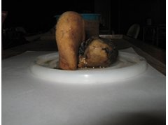
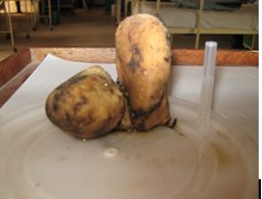
Fig 2: Upright cylindrical segment within the vagina; globular segment within the urinary bladder
Discussion
Obstetric trauma secondary to prolonged labour remains the commonest cause of VVF in developing countries1,2, and majority of the fistulae are due to obstructed labour at the first pregnancies unattended to by an appropriate healthcare professional 3. In Nigeria, Hilton et al4, found that 31% of cases were associated with first pregnancy.
A distressing complication of VVF repair is recurrence of the fistula which ranges from 19%4 to 37% 3 after primary repair. Urinary bladder calculi were found in 4 of 216 or 2% of cases of VVF evaluated radiologically by Lagundoye et al5, whilst it was found in 7% of VVF cases evaluated also radiologically by Akamaguna et al 6. This association was said to be uncommon7 and that the calculi tended to occur when any of the following conditions was present: high or supratrigonal fistula location, residual urine in the bladder, urinary contamination and a long history of disease. The other known predisposing and precipitating factors for vesical calculus formation in non VVF subjects were also applicable.
Our patient had a recurrent supratrigonal fistula of long duration, and the calculus incorporated nylon suture and vaginal tissue.
The calculus was progressing to form a complete cast of the bladder, vagina and the fistulous tract before the intervention, hence the dumb-bell shape. The shape and size of this stone was remarkable.
This patients management was staged7 - removal of the stone at the first surgery and definitive repair of the fistula subsequently. Repair of the fistula could be undertaken in the same sitting8 if there was no significant oedema of the peri-fistula tissues unlike the situation in the index case which informed the decision to repair the fistula at the second stage operation.
Public enlightenment on the prevention of VVF, and prompt and effective repair by experienced surgeons would go a long way in prevention and ensuring good management outcome. The first attempt is the best chance at successful repair. Conclusion: We have presented a young lady with a large dumb-bell calculus forming across a VVF defect. Public enlightenment emphasizing the need for the first VVF repair to be done by an experienced surgeon in a proper setting is the best chance for a successful outcome.
Acknowledgement
We thank Dr Afrika Gasana for the French translation, and Dr Habib Ahmed for his guidance.
REFERENCES
- Mubeen RM, Naheed F, Anwar K. Management of vesicovaginal fistulae in urological context. J Coll Physicians Surg Pak 2007 Jan; 17(1): 28-31.
- Arrowsmith S, Hamlin EC, Wall LL. Obstructed labour injury complex: obstetric fistula formation and the multifactrial morbidity of maternal birth trauma in the developing world. Obstet Gynecol Surv 1996; 51:568-72.
- Husain A, Johnson K, Glowacki CA et al. Surgical management of complex obstetric fistula in Eritrea. J Womens Health (Larchmt). 2005; 14(9): 839-44.
- Hilton P, Ward A. Epidemiological and surgical aspects of urogenital fistulae: a review of 25years experience in southeast Nigeria. Int Urogynecol J Pelvic Floor Dysfunct 1998; 9(4): 189-94.
- Lagundoye SB, Bell D, Gill G, Ogunbode O. Urinary tract changes in obstetric vesicovaginal fisulae: a report of 216 cases studied by intravenous urography. Clin Radiol 1976 Oct; 27(4):531-39.
- Akamaguna AI, Odita JC, Ajabor LN, Okpere EE. Radiology of obstetric vesicovaginal fistula. Urol Radiol 1983; 5(4): 247-50.
- Dalela D, Goel A, Shakhwar SN , Singh KM. Vesical calculi with unrepaired vesicovaginal fistula: a clinical appraisal of an uncommon association. J Urol 2003 Dec; 170(6 pt 1): 2206-8.
- Patankar S, Dobhada S, Bhansali M. Vesicovaginal fistula with secondary vaginal stones. J Laparoendosc Adv Surg Tech A 2006 Aug; 16(4): 386-9.