HIDRADENITIS SUPPURATIVA - A CASE REPORT
Nnamonu MI
Department Of Surgery,
Jos University Teaching Hospital,
Jos.
E-mail: miknnamonu@yahoo.com
Grant support: None
Subvention: Aucun
Conflict of Interest: None
Abstract
Hidradenitis suppurativa is a chronic and disfiguring skin disease characterized by multiple abscesses and sinuses. Often it is not recognized early in this environment as a result of limited awareness of this condition. The author sought to review available knowledge on this condition and report a case currently being managed in a general surgery unit. Literature search was conducted using the Google search engine and a patient who presented to the authors unit is presented. In conclusion, hidradenitis suppurativa is a distressing disease. High index of suspicion and relevant investigations would improve early diagnosis; several options are available for treatment with good outcome depending on the stage of the disease. Key words:Hidradenitis suppurativa, apocrine glands, sinuses, abscesses, antibiotics, excision, skin grafting.
INTRODUCTION
Hidradenitis suppurativa is a skin disease that most commonly affects areas of the body bearing apocrine sweat glands or sebaceous glands, such as the axillae, breasts, inner thighs, groin and buttocks. It is a chronic inflammatory disease characterized by abscesses and sinus formation1. It is frequently misdiagnosed as boils. This results in delayed diagnosis, fragmented care, and progression to a chronic, disabling condition with abscess formation that has a profoundly negative impact on quality of life. Simple boils have a pointed appearance with shiny or purulent overlying skin. The lesions in hidradenitis appear more rounded and extend into the deeper layers of the dermis. Hidradenitis suppurativa ( from the Greek hidros, sweat and aden, glands) is also known as Verneuils disease or acne inversa, and occasionally is spelled hydradenitis2.
The cause is unknown but may involve a defect of terminal follicular epithelium3. It has traditionally been attributed to occlusion of the apocrine duct by a keratinous plug.2 Contributing factors include friction from axillary adipose tissue, sweat, heat, stress, tight clothing and hormonal and genetic components2. Hidradenitis suppurativa usually occurs after puberty and before age 40, hence the theory that there is a hormonal component to the pathogenesis. Furthermore, flare-ups have been associated with shorter menstrual cycles and longer duration of menstrual flow 2.There is a genetic component, as a study of 110 patients, reported 38% of the patients with a family history of this disease. This is thought to reflect a familial form with autosomal dominant inheritance2. Cigarette smoking is a recognized risk factor for both the development of hidradenitis suppurativa and the progression to a severe disease. Obesity is also a risk factor; the majority of patients are overweight, and both body-mass index and tobacco smoking have been directly correlated with the severity of this condition4,5 .
Various studies in Europe show that hidradenitis suppurativa is not a rare disease2,4 . A Danish study noted a prevalence of 4% in women2. A French community study in persons above 15 years old which was questionnaire-based showed a prevalence of 1% after 1 year 4. Another study of young adults (18 to 33 years of age) undergoing screening for sexually transmitted diseases showed a prevalence of up to 4%4 . Women are more frequently affected with a female: male ratio variously reported as from 3:1 to 4:12,4 . Women are also reported to be more likely to have genitofemoral hidradenitis suppurativa 4 .
Case Report
D.D a 54 year old male presented with multiple perianal swellings. The swellings had been on and off for a period of 30 years but this last crop had persisted for 16 months. There was no history of anal intercourse, instrumentation or radiation. He had no swellings in the axillae nor in the groins. His weight was 67 Kg and height 1.78m with a body mass index of =21. Physical examination showed multiple perianal swellings with several sinuses discharging pus (Figure 1).
Packed cell volume was 33%. The purulent discharge grew Staphylococcus aureus sensitive to ciprofloxacin.
The urea , electrolytes and creatinine were normal. Total leucocyte count was 14.3x10⁹/L with differentials of neutrophils 58%, lymphocytes 34%, monocytes 6% and eosinophils 2%. There was a left shift in the neutrophils. Platelets were adequate and the red blood cells showed stomatocytes+, anisocytosis+ and target cells+. HbsAg was reactive while Anti HCV and HIV screening were non reactive. Incisional biopsies were taken from the masses and the histologic finding was stratified squamous epithelium overlying a loose, oedematous stroma within which were seen sinus tracts surrounded by areas of fibrino-suppurative inflammation. There was no evidence of malignancy.
Patient had oral ciprofloxacin 500mg twice daily for five days according to the sensitivity report and is being managed with oral clindamycin 300mg twice daily and oral rifampicin 300mg twice daily for three months. He is showing good response.
Discussion:
The pathogenesis of hidradenitis suppurativa remains unclear. However, current understanding reports that it is a multifocal disease, in which atrophy of the sebaceous glands is followed by an early lymphocytic inflammation and hyperkeratosis of the pilosebaceous unit. This is followed by hair follicle destruction and granuloma formation4,5 . It is posited that subsequent healing processes produce scarring and sinus tract formation - processes that are exacerbated by the impaired mechanical integrity of the sinus tract epithelium 4 . Recent investigations report that the interleukin-12-interleukin 23 pathway and tumour necrosis factor alpha are involved in the pathogenesis of hidradenitis suppurativa, supporting the proposition that it is an immune or inflammatory disorder 4,5 .
Bacterial infection with Staphylococci, Escherischia coli and Streptococcus is considered as a secondary event in the pathogenesis5 . The diagnosis of hidradenitis suppurativa is generally made clinically. On physical examination, there are characteristic inflamed and non inflamed nodules with discharging abscesses in the axillary, inguinal and anogenital regions. The lesions occasionally extend beyond these areas and appear around the anus, on the buttock or on the breast in females. The nodules are located in the deeper dermis and are rounded rather than having the pointed, purulent appearance of simple boils2. Secondary lesions such as pyogenic granulomas in sinus tract openings, plaque-like induration, ropelike scars and giant, multiheaded comedones may also be found2.
To make a diagnosis of hidradenitis suppurativa , the patient usually has one of the following:
-active disease with one or more primary lesions in a designated site (axilla, groin or perianal region) plus a history of three or more discharging or painful lumps (abscesses) in designated sites since puberty. -inactive disease with a history of five or more draining or painful abscess-like lumps in designated sites since onset of puberty, in the absence of concurrent primary lesions6 .
Assessment of the severity of the disease is generally based on the Hurley staging system: 4
-Stage I: a few isolated lesions without scarring tracts. Long periods of remission may delay an immediate diagnosis of hidradenitis suppurativa -Stage II: recurrent abscesses or single or multiple widely separated lesions with sinus tracts and scarring. Remissions are rare at this stage. Most diagnosis of hidradenitis suppurativa occur at this stage, and referral to surgery is often indicated.
-Stage III: the most devastating clinical stage. It is characterized by diffuse, broad involvement with multiple interconnecting sinus tracts and abscesses across a broad area of the body. At this stage, scarring and oozing lesions are common. Because remission is unlikely, surgery is most often recommended, although other treatments may be considered and tried.6 About 1% of patients have progression to stage III disease.4
Biopsies and bacterial cultures are indicated only in atypical or refractory cases. Routine bacteriologic studies of the lesions in hidradenitis suppurativa are most frequently negative, although flares may be associated with superinfection involving a range of bacteria, including Staphylococcus aureus. If extensive surgery is planned, ultrasonography may help in the preoperative assessment by identifying subclinical extension of the lesions 4 . Rarely, the patient has a fever or is septic, or both. Should the patient have fever or signs of sepsis, further work-up such as blood count, blood cultures and chemistry should be considered 2 .
With limited data available on randomized clinical trials on the treatment of hidradenitis suppurativa, the choice among several reported treatments is generally guided by the results in case series as well as by clinical experience and availability of the various treatment modalities. Patients are advised to control their weight and refrain from using tobacco. Rubbing of affected skin should also be avoided 4.
Stage I disease is managed with topical therapy while systemic therapy is used in patients with severe disease. Surgery is recommended when there is scarring as medical management offers little benefit in this situation.
Topical therapy with Clindamycin 10mg/ml twice daily for three months has been reported to reduce the number of abscesses, nodules and pustules4,7 . Intralesional injections of glucocorticoids ( eg triamcinolone, 2 to 5 mg ) for individual lesions has been also reported, though this has not been well studied 4 .
In situations where topical treatment is insufficient, oral antibiotics (often those with anti-inflammatory and immunomodulatory properties) are commonly used. A small randomized trial which compared treatment using oral tetracycline at a dose of 500mg twice daily with topical clindamycin as described earlier showed no superiority of the oral therapy 4. Alternatively, combination antibiotic therapy is recommended. A combination of clindamycin and rifampicin, each at a dose of 300mg twice daily have been reported to reduce disease severity and produce significant improvement in quality of life.4,8 Other medications that have been reported as useful in include minocycline in combination with rifampicin 8. Anti-androgens are sometimes used in women. A regimen combines ethinyl estradiol given from days 5 through 25 of the menstrual cycle plus cyproterone acetate given on days 5 through 14.4 Another combines ethinyl estradiol and cyptoterone acetate given on cycle days 5 to 25. Both were reported to reduce the frequencies of abscesses, the quantity of discharge and the degree of pain and discomfort 4. Systemic immunosuppressive agents have been advocated recently for patients with severe disease. The tumour necrosis factor-alpha inhibitor Infliximab has been used with good results9. It is given at a dose of 5 mg per kilogram body weight at weeks 0, 2 and 6. Other agents that have been used with success include etanercept( 50mg twice weekly) and adalimumab (40 mg every two weeks after a loading dose of 80mg)4,10 .
Surgery is used in patients who have extensive scarring and in patients with stage III disease. Incision and drainage is discouraged as this usually leads to recurrence. Surgery may be limited involving localized excision of sinus tracts, cysts and roofs of abscesses with wounds left open to heal by secondary intention4. More extensive procedures involving wide excision of all hair-bearing skin in affected areas and subsequent wound cover by skin grafting gives better results but may necessitate severely mutilating procedures4,11 . The use of carbon dioxide lasers and neodymium : yttrium-aluminium-garnet laser has been reported in patients with severe disease with good results4 . Radiation therapy has also been reported but use is limited due to concern that on the long term, risks may outweigh benefits4,12 .
In conclusion, hidradenitis suppurativa is a distressing disease. High index of suspicion and relevant investigations would improve early diagnosis; several options are available for treatment with good outcome depending on the stage of the disease.
References
- Heidi N. Anus. In: Townsend CM, Beauchamp RD, Evers BM, Mattox KL ( Editors). Sabiston Textbook Of Surgery The Basis Of Modern Surgical Practice. 17th Edition Vol.2 2004, Elsevier, Philadelphia:1483-1512.
- Shah N. Hidradenitis Suppurativa: A Treatment Challenge. Am Fam Physician. 2005: 72(8): 1547-1552.
- Mundy LM, Doherty GM, Cobb JP. Inflammation, Infection, & Antimicrobial Therapy. In: Doherty GM, Way LW (Editors). Current Surgical Diagnosis & Treatment. 12th Edition. The Mc Graw Hill Companies. United States Of America. 2006: p 97-126.
- Jemec GBE. Hidradenitis Suppurativa. N Engl J Med. 2012; 366: 158-164.
- Yazdanyar S, Jemec GB. Hidradenitis suppurativa: a review of cause and treatment. Curr Opin Infect Dis. 2011; 24(2): 118-123.
- Beshara MA. Hidradenitis suppurativa: A Clinicians Tool for Early Diagnosis and Treatment. The Nurse Practitioner: The American Journal of Primary Health Care. 2010. 35(5): 24-28.
- Yazdanyar S, Jemec GB. Hidradenitis suppurativa: a review of cause and treatment. Curr Opin Infect Dis. 2011; 24(2): 118-123.
- Mendonca CO, Griffiths CE. Clindamycin and rifampicin combination therapy for hidradenitis suppurativa. Br J Dermatol. 2006; 154(5): 977-978.
- Haslund P, Lee RA, Jemec GB. Treatment of hidradenitis suppurativa with tumour necrosis factor-alpha inhibitors. Acta Derm Venereol. 2009; 89(6): 595-600.
- Van der Zee HH, Laman JD, de Ruiter L, Dik WA, Prens EP. Adalimumab (antitumour necrosis factor-alpha ) treatment of hidradenitis suppurativa ameliorates skin inflammation: an in situ and ex vivo study. British Journal Of Dermatology. 2012; 166(2): 298-305.
- Menderes A, Sunay O, Vayvada H, Yilmaz M. Surgical management of hidradenitis suppurativa. Int J Med Sci. 2010; 7(4): 240-247.
- Trombetta M, Werts ED, Parda D. The role of radiotherapy in the treatment of hidradenitis suppurativa: Case report and review of the literature. Dermatology Online Journal. 2010; 16(2):16
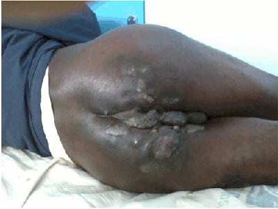
Figure 1: Patient with perianal hidradenitis suppurativa
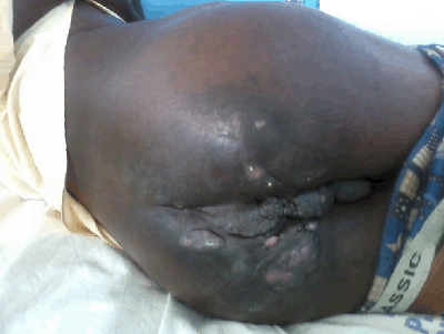
Figure 2: Pus discharging from sinuses in the same patient