UNUSUAL PRESENTATION OF FOREIGN BODY IN THE THIGH - A CASE REPORT
*Ezomike UO, Ituen MA, Ekpemo SC
Paediatric Surgery Unit, Department of Surgery, Federal Medical Centre, Umuahia, Nigeria.
E-mail: ezomikeuche@yahoo.com
Grant support: None
Subvention: Aucun
Conflict of Interest: None
Abstract
Foreign bodies in the thigh are uncommon. Rarer is the presence of a foreign body in the posterior compartment of the thigh following a wood puncture injury to the anterior thigh compartment. The purpose of this report is to highlight this unusual presentation. We present a six-year old boy presenting with a tumor-like mass in the posterior compartment of the right thigh thirteen months after a puncture injury to the anterior compartment of the right thigh. He sustained the injury while playing with a sharp wooden object. Part of the foreign body was expressed out while part of it was unknowingly left behind. The patient presented 13 months after with a mass in the posterior compartment of the thigh. He was promptly evaluated and he had exploration of the mass. A plain wooden foreign body was extracted which measured 6cm by 2cm. He made an uneventful postoperative recovery and has been followed up for more than twelve months without further symptoms. Adequate initial wound exploration with removal of all foreign bodies and necrotic tissues would have prevented this prolonged morbidity.
Wooden Foreign body, Posterior compartment of the thigh, Adequate Exploration
INTRODUCTION
Various foreign bodies have been extracted from the human thigh. They include wooden1, plastic, textile2 , metallic3, glass4 and ceramic5 materials. The patient is usually oblivious of the foreign bodies until they present with symptoms. They may access the thigh through accidental6 or iatrogenic means5.The tissue reaction to these foreign bodies varies according to the type of material and the patientís immune status. The manifestation may come at variable durations after the initial injury ranging from a few days to many years3,7. The presentation may be as thigh abscess, necrotizing fasciitis1, soft tissue sarcoma7, cyst6, pyogenic granuloma or tumor-like mass3. Ultrasonography, computed tomography and other imaging modalities have been used to identify such foreign bodies in the thigh. Treatment requires exploration and extraction of all the foreign bodies under the appropriate antibiotic and anti-tetanus covers.
Accidental access of wooden materials into the thigh resulting in a tumor-like mass, is hereby reported.
CASE REPORT
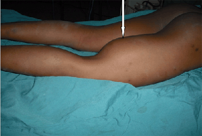
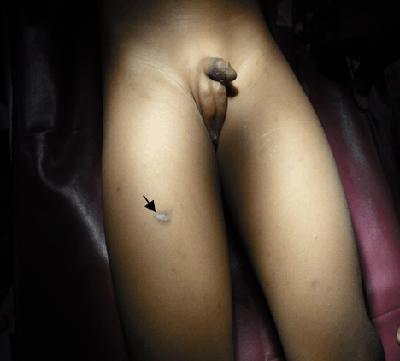
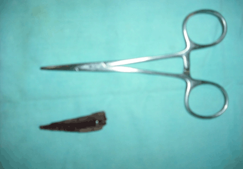
A 6-year old boy weighing 23kg presented with 7 months history of a painless swelling involving the posterior aspect of the right thigh. Six months earlier, he sustained a puncture injury to the anterior aspect of the same thigh caused by the sharp end of a piece of wood which the child was pushing along as a cart while playing. The impaled wood was promptly pulled out by his grandmother and he was taken to the hospital where efforts were made to express any remnant foreign body out of the wound. There was no formal wound irrigation or exploration but the wound was cleaned and dressed. He was given tetanus toxoid and antibiotics. The wound healed promptly. There was no restriction to the use of the limb as there was no pain. However, 13 months after the injury he presented to this facility with a mass in the right thigh. He was not pale and afebrile. There was a 10cm x 10cm non-tender mass on the posterior aspect of the right thigh which was fluctuant, not warm, and not attached to overlying skin. There was no neurovascular deficit of the affected limb. The hemoglobin level was 10.7g/dl with ESR of 63mm/hour and the blood film showed monocytosis. Ultrasonography revealed a well circumscribed cystic lesion with posterior acoustic enhancement and internal echoes harboring a linear echogenic substance suspected to be a foreign body in the subcutaneous plane. Plain radiograph of the thigh showed soft tissue swelling but no marginal irregularity and tissue planes were preserved; the femur was not involved. The patient was prepared and promptly operated. At surgery a thick-walled multiloculated cyst was found within the posterior compartment of the thigh containing serous non-offensive fluid. The intracystic septae were broken and a 6cm x 2cm arrow-shaped plain wooden material was removed. The cavity was cleaned out and the biopsy tissue of the wall was taken for histology while the serous fluid was cultured. A close tube drain was inserted, the wound was closed and dressed. Post-operative recovery was uneventful. The patient has been followed up for more than one year, and has remained without symptoms.
DISCUSSION
The presence of a wooden foreign body in the posterior thigh compartment of a child following a puncture injury to the anterior compartment is not common in literature. In this case, inadequate initial wound care which did not include wound exploration was the cause of the morbidity. Proper wound exploration and irrigation under anesthesia would have revealed the remnant foreign body during the initial management and would have prevented this late presentation. Efforts to squeeze out any intra-lesional foreign body out of the wound during initial management may have encouraged migration posteriorly. Moreover, pulling out an impaled foreign body blindly without wound exploration could be disastrous as the foreign body could lacerate major blood vessels and nerves with severe consequences.
The fact that the wood was not painted may have reduced the inflammatory reaction to it. Also it was not complicated by abscesses, necrotizing fasciitis or tenderness which when present would be associated with complaints of pain1. The arrow-shape of the foreign body and attempts at squeezing it out may have enhanced the migration to the posterior compartment. It is also unusual for the initial wound to have healed uneventfully with an unsterile foreign body in its depths.
Tissue reaction to foreign body is variable. Deep tissue abscess formation with or without necrotizing fasciitis was a more likely reaction here considering the dirty environment and injuring agent1. However the foreign body was encased in a thick-walled fibrous cavity with no obvious clinical evidence of infection. This may be due to initial antibiotics given to the patient. Another complication of foreign bodies in tissues is malignant transformation such as angiosarcoma7. Fungal masses have been formed around wooden foreign bodies forming a phaeomycotic mass6. Inert substances like bullets elicit minimal or no reaction hence the teaching that such should be left unless they are symptomatic.
Plain radiography did not reveal the foreign body. This corroborated other studies which observed low sensitivity of plain radiography in detecting radiolucent foreign bodies5.Ultrasonography detected the foreign body in this case as were the cases reported by Jacobson8 and Bushberg 9. However the foreign body was wrongly localized by ultrasonography to the subcutaneous plane when it was actually in the intermuscular plane; this corroborated with the notion that ultrasonography was operator-dependent. Computed tomography scan would have given a more accurate finding but was not available in our hospital when the patient presented.
It has also been shown that apart from direct impalement, foreign bodies have also migrated to the thigh by such routes as transperitoneal migration 10, transcutaneous penetration of wooden splinters6 and as inadvertently retained surgical sponges.2
In conclusion, impalement foreign bodies are better explored promptly in order to extract the entire foreign body on the day of injury under anesthesia and with the appropriate antibiotics and anti-tetanus cover.
References
- Yanay O, Vaughan DJ, Diab M, Brownstein D, Brogan TV. Retained wooden foreign body in a childís thigh complicated by severe necrotizing fasciitis: A case report and discussion of imaging modalities for early diagnosis. Paediatr Emerg Care.2001 Oct;17(5):354-5.
- Puri A, Anchan C, Jambhekar NA, Agarwal MG, Badwe RA. Recurrent gossypiboma in the thigh.Skeletal Radiol.2007 Jun;36 Suppl 1.S95-100.
- Kookkinakis M, Rajeev A, Newby M, Graham D. An unusual presentation of a giant tumour-like lesion of the thigh.ActaOrthop Belg.2008 Jun;74(3):421-3.
- Reynier C , Dubost JJ, Marquet C, Lhoste A, Guillon R, Sauvezie B, Michel JL : Glass foreign body of the posterior part of the right thigh. JRadiol 2000 Aug;81(8):902-3.
- Ando A, Hatori M, Hagiwara , Isefuku S, Itoi E: Imaging features of foreign body granuloma in the lower extremities mimicking a soft tissue neoplasm. Uppsala Journal of Medical Sciences.2009;114:46-51.
- Iwatsu T, Miyaji M . Phaeomycotic cyst. A case with a lesion containing a wooden splinter .Arch Dermatol.1984 Sept;120(9):1209-11.
- Jennings TA, PetersonL, AxiotisCA, FriedlaenderGE,CookeRA,RosaiJ.Angiosarcoma associated with foreign body material: A report of three cases. Cancer.1988 Dec 1;62(11):2436-44.
- Jacobson JA, Powell A, Craig JG, Bouffard JA, VanHolsbeeck MT: Wooden foreign bodies in soft tissue: detection at ultrasound. Radiology.1998 Jan;206(1):45-8.
- Bushberg JT, Seibert JA, Leidholdt EMJ, Boone JM: The Essential Physics of Medical Imaging, 2nd ed. Philadelphia: PA,Lippincott Williams&Wilkins,2001.
- Leelouche N, Ayoub N, Bruneel F, Mignon F, Troche G, Boisrenault P, Bedos JP: Thigh cellulitis caused by toothpick ingestion. Intensive Care Med.2003 Apr;29(4):662-3.

Figure 1: Tumor-like mass on the posterior aspect of right thigh

Figure 2: Arrow showing scar at the puncture site on the anterior aspect of the right thigh

Figure 3: Retrieved arrow-shaped wooden foreign body